


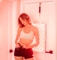


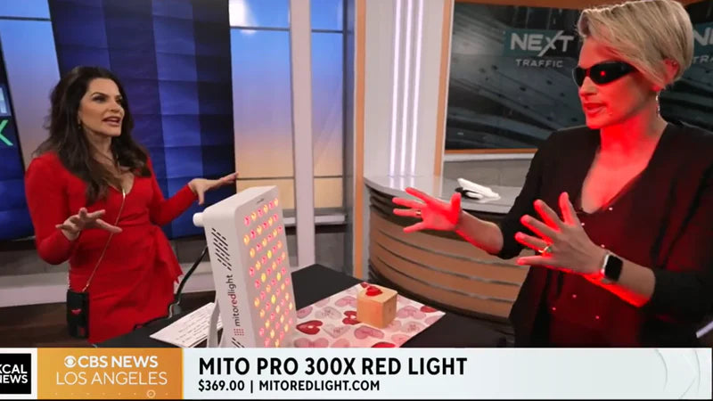

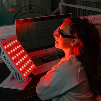


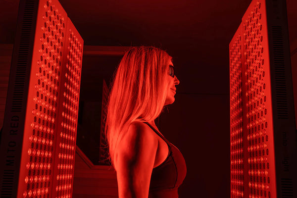
Panel Series
Photobiomodulation (PBM) research suggests red and near-infrared light therapy may accelerate muscle recovery by increasing mitochondrial ATP production, reducing exercise-induced oxidative stress, and attenuating inflammatory signaling in muscle tissue. Multiple meta-analyses and human RCTs have documented reduced delayed-onset muscle soreness (DOMS), lower creatine kinase levels, and faster return-to-performance following structured PBM protocols.
New to red light therapy? Read our introduction to red light therapy before diving into the muscle recovery research below.
Reviewed for scientific accuracy by Dr. Alexis Cowan, PhD in Molecular Biology (Princeton University), who specializes in mitochondrial function and photobiomodulation research.
Red Light Therapy and Muscle Recovery: The Direct Answer
Red light therapy — also called photobiomodulation, or PBM — refers to the application of specific wavelengths of non-thermal red and near-infrared light, typically between 630nm and 900nm, to stimulate biological function at the cellular level. In the context of muscle recovery, research suggests it may reduce the severity of delayed-onset muscle soreness (DOMS), lower circulating markers of muscle damage such as creatine kinase (CK) and lactate dehydrogenase (LDH), decrease inflammatory cytokines following exercise, and support faster return to peak performance between training sessions.
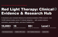
The evidence base is substantial — the Muscle category in Mito Red Light's photobiomodulation research evidence hub contains over 398 studies including 18 meta-analyses, 9 systematic reviews, and 178 human trials. The findings are consistent enough that a 2016 review in the Journal of Biophotonics by Ferraresi, Huang, and Hamblin raised the question of whether PBM should be regulated in competitive athletics due to the potential performance advantage it may confer.
That said, the evidence is not uniformly positive. Several well-designed RCTs — particularly in already-trained athletes and in high-intensity anaerobic protocols — show no significant benefit over sham. The honest picture is that PBM appears most effective for recovery and for pre-exercise muscle protection, with more mixed results for acute performance enhancement, and with effect sizes that vary meaningfully by protocol, population, and application timing.
For a complete indexed library of peer-reviewed PBM clinical studies organized by health category, Mito Red Light maintains a clinical research evidence hub that includes the muscle recovery and sports performance evidence in full detail.
Why Muscles Need Recovery: The Biology of Exercise-Induced Damage
Understanding why red light therapy may support muscle recovery requires understanding what exercise actually does to muscle tissue at the cellular level — because recovery is not simply a matter of rest, it is an active, energy-intensive biological process.
When muscle fibers are subjected to mechanical load — especially eccentric contractions, where the muscle lengthens under tension as in the lowering phase of a squat or the landing phase of a jump — the physical stress causes disruption of the contractile proteins (actin and myosin), damage to the sarcolemma (cell membrane), and release of calcium into the cell interior. This initiates a cascade of events: inflammatory cells infiltrate the damaged tissue, reactive oxygen species (ROS) accumulate, and the affected muscle cells begin a repair and remodeling process.
The delayed soreness most people experience 24–72 hours after an unfamiliar or intense workout — DOMS — is a product of this inflammatory and repair process, not of lactic acid accumulation as commonly believed. Lactate clears from muscle within hours; DOMS peaks a day or more later as the inflammatory cascade progresses and mechanoreceptors in the damaged tissue become sensitized.
The energy cost of this repair process is substantial. Satellite cells (muscle stem cells) must proliferate and differentiate to rebuild damaged fibers, protein synthesis must ramp up to replace disrupted contractile elements, and the inflammatory response must be resolved before the muscle can function optimally again. Anything that supports mitochondrial energy production, modulates the inflammatory response, or reduces the initial extent of oxidative damage will, in theory, accelerate this process.
This is precisely the biological terrain where photobiomodulation operates. For a detailed explanation of the cellular mechanism — including cytochrome c oxidase (CCO) activation, ATP production, and secondary signaling cascades — see how red and near-infrared light trigger cellular response at the mitochondrial level.
"The muscle recovery application makes mechanistic sense when you look at what the exercise-damage cascade actually requires. You need ATP for the repair process, you need controlled ROS signaling rather than runaway oxidative stress, and you need the inflammatory response to resolve on schedule rather than persist. Near-infrared light at the wavelengths that activate cytochrome c oxidase addresses all three of those directly — it improves mitochondrial efficiency, modulates ROS through secondary signaling, and has well-documented effects on inflammatory mediators. The question isn't really whether the mechanism is plausible, it clearly is, it's about optimising the dose and timing to translate that into consistent clinical outcomes."
— Dr. Alexis Cowan, PhD, Molecular Biology (Princeton University), Scientific Advisor, Mito Red Light
What the Clinical Evidence Shows
The muscle recovery evidence base is among the most developed in the photobiomodulation literature. The following sections cover the current state of evidence organized by outcome type, from the highest-quality systematic reviews down to recent human RCTs.
Meta-Analyses and Systematic Reviews: The Highest-Level Evidence
Multiple independent meta-analyses have now examined the PBM and muscle recovery literature, and their conclusions are broadly consistent.
A 2022 meta-analysis by Luo et al., published in Sports Health, analyzed 24 randomized controlled trials examining the effects of low-level laser therapy on muscular performance and soreness recovery in athletes. The review found consistent reductions in post-exercise soreness and documented improvements in at least three exercise-related biomarkers following PBM. This meta-analysis is the most frequently cited recent synthesis of the field and establishes a meaningful evidence base across diverse athletic populations and exercise types.
A 2022 meta-analysis by De Marchi et al., published in Antioxidants (Basel), specifically examined whether PBM minimizes exercise-induced oxidative stress. The systematic review found evidence supporting this effect, consistent with the proposed mechanism of CCO-mediated ROS modulation. Oxidative stress is a primary driver of both DOMS severity and the duration of the recovery window, making this finding mechanistically important.
A 2022 meta-analysis by Dutra et al. published in Sports Medicine took a particularly rigorous approach, attempting to deconstruct the ergogenic effects of PBM by exercise modality. The analysis found positive effects for single-joint muscle endurance and cycling time-to-exhaustion, while finding no significant benefit for running performance, swimming, cycling sprints, or muscle strength — a finding that underscores the protocol-specificity and population-specificity documented throughout this literature.
A 2025 meta-analysis by Qiu et al. published in Sports Health specifically examined volleyball and football (soccer) players across randomized controlled trials. The analysis found that PBM improved muscle performance in these team-sport populations, adding to the growing sport-specific evidence base.
A 2025 systematic review by Álvarez-Martínez and Borden, published in Lasers in Medical Science, examined whole-body PBM specifically for exercise performance and recovery — a methodologically important distinction, as most earlier trials used localized application. The review found evidence supporting whole-body protocols for both performance and recovery outcomes, which is relevant for those using full-panel devices rather than targeted probes.
Reducing Muscle Damage: Pre-Exercise Application
One of the most consistently supported findings in the PBM sports literature is that application before exercise reduces the extent of muscle damage that occurs during that exercise. This appears to work through a preconditioning mechanism — PBM upregulates antioxidant defenses and supports mitochondrial function before the oxidative challenge of exercise begins, reducing the severity of the initial damage cascade.
A 2023 RCT by Crow et al., published in the Journal of Athletic Training, enrolled athletes and applied PBM before strenuous resistance exercise targeting the shoulder. In the final blocks of exercise (sets 9–12), all performance measures in the active PBM group were 6.2% to 10% greater than sham values (p < 0.02 to 0.001). The effect was most pronounced as cumulative fatigue accumulated — consistent with PBM's proposed role in maintaining mitochondrial function and ATP availability during sustained effort.
A 2024 study by Santos et al. published in Frontiers in Sports and Active Living combined warm-up with PBM and measured performance on the Yo-Yo Intermittent Recovery Level 1 test. The warm-up + PBM group achieved 440.0 ± 59.0 m compared to 353.3 ± 94.7 m in the warm-up + placebo group (p = 0.02) — a meaningful difference in a sport-specific endurance test.
A 2019 RCT by Dornelles et al., published in Lasers in Medical Science, used a randomized, crossover, double-blinded, placebo-controlled design and found that PBM applied before a soccer match attenuated hamstring muscle fatigue — a finding the authors identified as supporting PBM as a promising tool for hamstring strain prevention in team sport athletes.
Reducing DOMS and Accelerating Post-Exercise Recovery
Post-exercise application of PBM has been studied extensively for its ability to reduce DOMS severity and accelerate recovery of muscle function. Several mechanisms are proposed: reduced inflammatory cytokine signaling (particularly IL-6, TNF-alpha, and IL-1β), improved lymphatic drainage, faster clearance of metabolic byproducts, and enhanced mitochondrial support for the active repair process.
A 2022 RCT by Pinto et al., published in Oxidative Medicine and Cellular Longevity, followed CrossFit athletes receiving PBM combined with a static magnetic field. The treatment group showed enhanced performance in functional tests, decreased levels of biochemical markers of muscle damage and inflammation, decreased oxidative stress, and increased antioxidant activity compared to placebo. This study is notable for its use of a well-validated real-world athletic population and its inclusion of both performance and biomarker outcomes.
A 2023 RCT by Chen et al., published in Medicine & Science in Sports & Exercise, examined post-contraction recovery with ischemic preconditioning. At the follow-up measurement, the laser group demonstrated a higher normalized MVC (86.22 ± 12.59%) compared to the sham group (71.70 ± 13.56%; p = .001) — a statistically and clinically meaningful difference in contractile function recovery.
A 2023 study by Lanferdini et al., published in the Journal of Functional Morphology and Kinesiology, applied PBM before successive time-to-exhaustion cycling tests and found approximately 10–12% improvement in performance in the first and second tests, along with improved VO₂ and deoxyhaemoglobin (HHb) kinetics — physiological markers indicating more efficient oxygen utilization in muscle tissue.
A 2014 study by Borges et al., published in Lasers in Medical Science, used a randomized, double-blinded, placebo-controlled design and demonstrated that a single LED therapy session improved muscle soreness, preserved muscle strength, and reduced range-of-motion impairments caused by exercise, with effects persisting for up to 96 hours post-exercise.
A 2016 RCT by Pinto et al., published in the Journal of Strength and Conditioning Research, followed high-level rugby players in a randomized, crossover, double-blind, placebo-controlled study and found that PBM meaningfully accelerated post-match muscle recovery — an important finding in a contact sport population where recovery between fixtures is a primary performance constraint.
The full library of muscle recovery and sports performance research is accessible through Mito Red Light's searchable database of over 9,500 peer-reviewed PBM studies.
Creatine Kinase and Inflammatory Biomarkers
One of the most objective ways to measure muscle damage is through blood biomarkers. Creatine kinase (CK) leaks from damaged muscle cells into the bloodstream; elevated CK is a direct proxy for the degree of muscle fiber disruption following exercise. Lactate dehydrogenase (LDH) is another muscle damage marker. Inflammatory cytokines including IL-6, TNF-alpha, and C-reactive protein (CRP) reflect the extent and duration of the post-exercise inflammatory response.
Multiple RCTs in the existing literature have examined PBM and post-exercise CK and LDH levels compared to sham, with a number of studies reporting reductions in these markers following treatment.
A 2016 double-blind, randomized, placebo-controlled study by Zagatto et al., published in Lasers in Medical Science, used water polo athletes and found that infrared light treatment was associated with reductions in CK over time within the treatment group. The authors reported small-to-moderate effects on inflammatory and muscle damage markers overall, though between-group differences for inflammatory markers including IL-1β and TNF-alpha did not reach statistical significance. The sport-specific population and validated biomarker outcomes give this study contextual value in the PBM recovery literature.
A 2022 meta-analysis by Yang et al. (published in Photochemistry and Photobiology) specifically analyzed inflammatory factors during skeletal muscle regeneration across animal studies, finding consistent evidence for PBM-mediated reduction in pro-inflammatory cytokines — consistent with some human biomarker findings but providing mechanistic depth from controlled preclinical models.
For the muscle damage biomarker and inflammation evidence reviewed in a clinical context, the clinical evidence on PBM and muscle recovery and performance provides a structured review organized by study type.
Where the Evidence Is Mixed: Honest Limitations
Calibrated confidence is essential in evaluating this literature. Not all studies show benefit, and the pattern of null findings is informative.
Several well-designed RCTs have found no significant benefit from PBM in specific contexts. A 2025 RCT by Caseiro-Filho et al., published in Lasers in Medical Science, found PBM "ineffective in eliciting performance enhancements" in a resistance training program for lower limbs in healthy individuals — with the authors noting that "divergent parameters evince equivocal efficacy," pointing to dose and irradiance specificity as key variables. A 2025 RCT by Borim et al. found no improvement in muscle endurance, power, or strength in female futsal players using LED-based PBM.
A 2024 RCT by do Nascimento et al., published in Lasers in Medical Science, tested acute PBM across multiple doses (300, 900, and 1,260 J) before a 5 km running trial in recreational runners and found none of the doses improved endurance performance or perceived exertion.
A 2023 RCT by Forsey et al. found that whole-body PBM applied before maximal anaerobic cycling improved post-exercise recovery but did not improve performance during the exercise itself — an important distinction that helps define where in the exercise window PBM's effects are strongest.
The pattern that emerges from the null findings is coherent: PBM appears most effective for recovery-oriented outcomes (DOMS reduction, faster return of force output, lower biomarkers of damage) and for pre-exercise muscle protection, with less consistent evidence for acute performance enhancement in already well-trained athletes or in high-intensity anaerobic protocols of short duration.
A 2022 study by Machado et al. specifically found no additional benefit when PBM was added to a six-week training program in previously trained individuals — suggesting training status and adaptation state modulate the response.
This honest picture is more useful for practitioners than a uniformly positive summary would be. The evidence supports PBM as a recovery and injury-prevention tool, with performance effects that are real but context-dependent and protocol-sensitive.
How Red Light Therapy Works in Muscle Tissue: Proposed Mechanisms
The following mechanisms have been proposed in the PBM literature to explain its effects on muscle tissue. They are presented as hypotheses supported by varying degrees of evidence, not as established facts.
CCO Activation and Mitochondrial ATP Production
The primary proposed mechanism for PBM in muscle tissue is the same as in other tissues: activation of cytochrome c oxidase (CCO), the terminal enzyme of the mitochondrial electron transport chain. CCO has well-characterized absorption peaks in the red and near-infrared spectrum (approximately 660nm and 830nm), and photon absorption at these wavelengths is proposed to enhance electron transfer, increase proton gradient, and ultimately increase ATP synthesis. In actively recovering muscle, where energy demand for protein synthesis and repair processes is high, enhanced mitochondrial efficiency is hypothesized to accelerate the timeline of functional recovery.
ROS Modulation
Reactive oxygen species (ROS) play a dual role in muscle adaptation: at low levels, they are necessary signaling molecules that trigger adaptation responses; at high levels following intense exercise, they contribute to oxidative damage and delayed recovery. PBM is proposed to modulate this balance — supporting the low-level ROS signaling needed for adaptation while attenuating the excess oxidative load that extends the damage-repair timeline. The De Marchi (2022) meta-analysis specifically examined this mechanism and found supporting evidence across the human and preclinical literature.
Anti-Inflammatory Signaling
In the existing PBM literature, photobiomodulation has been associated with effects on inflammatory mediators including NF-κB (a master regulator of inflammatory gene expression), prostaglandin E2, IL-1β, IL-6, and TNF-alpha. In the muscle recovery context, this is proposed to accelerate resolution of the post-exercise inflammatory phase — moving the tissue faster from the inflammatory/damage phase into the regeneration and remodeling phase without eliminating the inflammatory response entirely (which would impair adaptation). This is a critical nuance: the goal is not to suppress inflammation completely, which would interfere with muscle adaptation, but to normalize its duration and intensity.
Nitric Oxide Release
PBM has been described to cause release of nitric oxide (NO) from its bound forms in mitochondria and hemoglobin. NO is a potent vasodilator, and increased local blood flow following PBM application may enhance delivery of oxygen and nutrients to recovering muscle tissue while supporting clearance of metabolic byproducts. Improved microcirculation is particularly relevant in the early recovery phase when metabolic demands in damaged tissue are high.
Satellite Cell Activation
Preclinical studies suggest PBM may activate muscle satellite cells — the stem cells responsible for regenerating damaged muscle fibers. A 2021 narrative review published in Research, Society and Development analyzed evidence for PBM in muscle atrophy and found it may stimulate muscle cell growth, enhance formation of muscle fibers, and support cellular maintenance processes. These findings are primarily from animal models and in vitro work, and their direct applicability to exercise recovery in healthy humans requires further investigation. For the full evidence on PBM and cellular energy metabolism, the clinical evidence on PBM and mitochondrial function covers the mechanistic research in detail.
Practical Protocol: How to Use Red Light Therapy for Muscle Recovery
The following reflects the approaches used in published human studies and the parameters that have shown the most consistent results. This is educational information, not medical advice. Those with musculoskeletal injuries, relevant medical conditions, or questions about specific protocols should consult a healthcare provider.
Pre-Exercise vs. Post-Exercise: Does Timing Matter?
The evidence consistently supports pre-exercise application for muscle protection and performance effects, with post-exercise application most relevant for DOMS reduction and accelerating recovery between sessions. Several studies have tested both timing windows directly.
A 2024 RCT by da Costa Santos et al., published in the Journal of Bodywork and Movement Therapies, compared pre-exercise versus post-exercise PBM application in rugby athletes at 850nm. The pre-exercise group showed improved performance on the first Yo-Yo test compared to both post-exercise and placebo groups — suggesting pre-exercise timing has a specific advantage for acute performance effects. Recovery biomarkers (CK and lactate) did not differ significantly between groups, pointing to timing specificity for different outcomes.
The practical implication is that pre-exercise application (typically 5–30 minutes before training or competition) may be the more critical window for those seeking performance and muscle protection effects, while post-exercise application may be more appropriate as part of a recovery routine.
Wavelength Reference for Muscle Recovery
| Wavelength | Tissue Penetration | Primary Mechanism in Muscle | Studies Using This Wavelength |
|---|---|---|---|
| 630–660nm (Red) | ~1–2cm | Superficial muscle tissue, CCO activation, anti-inflammatory | Flores (2023), Rodrigues (2013) |
| 808nm (NIR) | ~3–5cm | Deep muscle penetration, strong CCO affinity, mitochondrial ATP | Leal Junior (2009), Azuma (2021) |
| 830nm (NIR) | ~3–5cm | Deep NIR, anti-inflammatory, fatigue reduction | Leal Junior (2009), Crow (2023) |
| 850nm (NIR) | ~3–5cm | Deep NIR, broad muscle recovery and fatigue effects | da Costa Santos (2024), Chen (2023) |
| 904nm (NIR) | ~5–7cm | Deepest penetration, reaches large muscle bellies | Silva (2025) |
Near-infrared wavelengths (808–850nm) dominate the muscle recovery literature because their greater tissue penetration reaches the belly of most skeletal muscles — a prerequisite for meaningful photobiomodulation of deep muscle tissue. Combination panels delivering both 660nm red and 850nm NIR cover both the surface tissue effects and the deeper muscle targets used in the majority of clinical protocols.
The MitoPRO X Series delivers both 660nm and 850nm at clinical irradiance levels — the same dual-wavelength range that appears across much of the human PBM literature on musculoskeletal recovery. For detailed wavelength-specific dosing parameters and penetration depth data, see Mito Red Light's wavelength and dosing reference.
Session Parameters from the Clinical Literature
PBM protocols in the muscle recovery literature vary considerably. The following reflects the range of approaches used across studies that showed positive outcomes:
- Dose (fluence): Most positive studies have used 10–50 J/cm² applied to each muscle group. A 2020 review in the Journal of Sport Rehabilitation specifically examining soccer athletes found doses between 10 and 50 J provided the greatest effect. Higher doses have shown equivocal results in some studies — suggesting an optimal dose window rather than a "more is better" relationship.
- Session duration: Typically 5–20 minutes for targeted application per muscle group; whole-body protocols in panel studies range from 10–20 minutes.
- Frequency: Most RCTs in recovery contexts apply PBM immediately before or after each training session. Daily use during high-training-load periods is consistent with the protocols showing the most robust results.
- Distance: Device-to-skin distance affects irradiance significantly. Most clinical protocols use contact or near-contact application for targeted probes; panels are typically used at 15–30cm per manufacturer protocol.
For a comprehensive guide to device selection including irradiance specifications, coverage area, and what to look for in a panel for athletic use, the red light therapy buyer's guide covers the key specifications and common buyer mistakes.
Red Light Therapy Within a Broader Recovery Strategy
PBM works most effectively as part of a comprehensive recovery approach rather than in isolation. The following recovery modalities have their own evidence base and interact with PBM's effects.
Sleep is the primary recovery tool for muscle tissue. Growth hormone release, protein synthesis upregulation, and systemic anti-inflammatory processes are all concentrated in deep sleep phases. A 2011 study published in Medical Hypotheses documented the endocrinological and molecular basis for sleep's role in muscle recovery, particularly its influence on protein synthesis. PBM may complement sleep-related recovery, but it does not substitute for adequate sleep duration and quality.
Protein intake timing matters for muscle repair. The building blocks for replacing damaged contractile proteins must be available during the repair window. Post-exercise protein intake, particularly within the period following training, is well-supported in the sports nutrition literature for supporting muscle protein synthesis — the process PBM's mitochondrial support may help energize.
Active recovery — light movement, walking, or low-intensity cycling — supports lymphatic drainage and blood flow without creating additional mechanical stress. Several studies have combined PBM with active recovery modalities. A 2025 systematic review by Canez et al. in the Journal of Bodywork and Movement Therapies compared PBM, intermittent pneumatic compression, and neuromuscular electrical stimulation for muscle recovery, finding complementary rather than competing effects — supporting the use of PBM as one tool within a multimodal recovery protocol.
Hydration is foundational. Muscle cells require adequate hydration for enzymatic function, electrolyte balance, and cellular repair. A study in the Journal of Human Kinetics confirmed that hydration improves both athletic performance and post-exercise recovery. This is not modulated by PBM and must be addressed independently.
For an overview of how red light therapy compares to professional treatment settings and what at-home protocols can realistically achieve, the at-home vs. clinic red light therapy comparison covers the practical differences.
Red Light Therapy for Muscle Injuries: What the Evidence Supports
Beyond routine post-exercise recovery, PBM has been studied in the context of acute muscle injuries — strains, contusions, and sports injuries requiring structured rehabilitation.
A 2023 RCT by Wells et al., published in the Journal of Sport Rehabilitation, applied pulsed red and blue PBM to thigh contusions in athletes. The active PBM group showed significantly greater quadriceps peak torque at 180°/s (p = 0.030) and average power at both 60°/s and 180°/s compared to placebo. For a common and performance-limiting sports injury type, this is clinically relevant evidence.
A 2024 meta-analysis by Morgan et al., published in the Journal of Strength and Conditioning Research, examined the effects of PBM specifically on pain reduction and return-to-play timelines in injured athletes across RCTs. The analysis found meaningful effects on both outcomes — relevant for the practical question of whether PBM shortens the time from injury to competition-readiness.
A 2024 meta-analysis by Alayat et al., published in Lasers in Medical Science, specifically examined PBM effectiveness for ankle sprains — one of the most common acute sports injuries — and found supporting evidence for its use in this population.
A 2013 study by Morimoto et al., published in Laser Therapy, examined 41 sports injury patients in a clinical setting and documented a 65.9% effectiveness rate across all sports injury types, with higher rates for specific conditions including jumper's knee, tennis elbow, and Achilles tendinitis. While this is an older study with methodological limitations, the clinical breadth of conditions studied gives it contextual value.
Anyone with an acute muscle injury should consult a sports medicine physician or physical therapist before relying on any single modality as a primary intervention. PBM may support recovery within a supervised rehabilitation plan, but it does not replace assessment and treatment of the underlying injury. For the full indexed evidence on PBM and musculoskeletal conditions, the clinical evidence on inflammation and pain covers the related injury and pain literature.
Frequently Asked Questions
Does red light therapy actually help muscle recovery?
Multiple meta-analyses and human RCTs support the use of PBM for muscle recovery outcomes including reduced DOMS, lower CK and inflammatory markers, and faster return of force output. A 2022 meta-analysis in Sports Health (Luo et al.) reviewed 24 RCTs and found consistent post-exercise soreness reduction and biomarker improvements. However, the evidence is not uniformly positive — several well-designed trials show no benefit in already well-trained athletes or in high-intensity anaerobic protocols. The strongest evidence supports pre-exercise application for muscle protection and post-exercise application for DOMS reduction.
Should I use red light therapy before or after a workout?
Both windows have evidence. Pre-exercise application (5–30 minutes before training) is supported for reducing muscle damage during exercise and potentially extending time to fatigue. Post-exercise application is most relevant for reducing DOMS severity and accelerating functional recovery between sessions. A 2024 RCT in rugby athletes (da Costa Santos et al.) found pre-exercise application specifically improved performance in the first high-intensity test, while post-exercise effects on biomarkers were less differentiated. For athletes primarily concerned with performance preservation across a training block, pre-exercise timing may be the priority.
What wavelength is best for muscle recovery?
Near-infrared wavelengths in the 808–850nm range dominate the muscle recovery literature because they penetrate 3–5cm into tissue — deep enough to reach the belly of most skeletal muscles. Red wavelengths (630–660nm) are primarily absorbed in surface tissue and are less relevant for deep muscle applications, though they may contribute to surface-level anti-inflammatory effects. Most research protocols use NIR, with 808nm and 830nm being the most commonly studied wavelengths for muscle outcomes specifically.
How long does it take to see results from red light therapy for muscle recovery?
Single-session effects on DOMS and soreness have been documented in RCTs — a 2014 study (Borges et al.) found that a single LED therapy session improved muscle soreness and preserved strength with effects persisting for up to 96 hours post-exercise. For cumulative effects on training adaptation and injury prevention, studies using multi-week protocols consistently show more robust results than single-session designs. The honest answer is that some recovery benefit may be apparent immediately, while the full benefit of consistent use over a training block takes weeks to accumulate.
Can red light therapy help with muscle strains and sports injuries?
Preliminary evidence suggests PBM may support recovery from muscle injuries including strains and contusions. A 2024 meta-analysis (Morgan et al.) found meaningful effects on pain and return-to-play timelines in injured athletes. A 2023 RCT (Wells et al.) documented improved quadriceps peak torque in thigh contusion patients receiving PBM versus placebo. PBM is not a substitute for medical evaluation of acute injuries — anyone with a significant muscle strain or sports injury should be assessed by a sports medicine professional before relying on any single recovery modality.
References
- Luo WT, Lee CJ, Tam KW, Huang TW. (2022). Effects of Low-Level Laser Therapy on Muscular Performance and Soreness Recovery in Athletes: A Meta-analysis of Randomized Controlled Trials. Sports Health, 14(5), 687–693. PMID 34428975
- De Marchi T, et al. (2022). Can Photobiomodulation Therapy (PBMT) Minimize Exercise-Induced Oxidative Stress? A Systematic Review and Meta-Analysis. Antioxidants (Basel). PMID 36139746
- Dutra YM, et al. (2022). Deconstructing the Ergogenic Effects of Photobiomodulation: A Systematic Review and Meta-analysis of its Efficacy in Improving Mode-Specific Exercise Performance. Sports Medicine. PMID 35802348
- Qiu Y, et al. (2025). The Effect of Photobiomodulation Therapy on Muscle Performance in Volleyball and Football Players: A Meta-Analysis of Randomized Controlled Trials. Sports Health. PMID pending verification
- Álvarez-Martínez D, Borden A. (2025). A systematic review on whole-body photobiomodulation for exercise performance and recovery. Lasers in Medical Science. PMID pending verification
- Crow JA, et al. (2023). Therapeutic Photobiomodulation Before Strenuous Exercise Attenuates Shoulder Muscle Fatigue. Journal of Athletic Training. PMID 38015822
- Santos HH, et al. (2024). Innovative integration: optimizing performance through warm-up and photobiomodulation in high-intensity test. Frontiers in Sports and Active Living. PMID pending verification
- Dornelles MP, et al. (2019). Photobiomodulation therapy as a tool to prevent hamstring strain injuries by reducing soccer-induced fatigue on hamstring muscles. Lasers in Medical Science, 34(6), 1177–1184. PMID 30607719
- Pinto HD, et al. (2022). Photobiomodulation Therapy Combined with a Static Magnetic Field Applied in Different Moments Enhances Performance and Accelerates Muscle Recovery in CrossFit Athletes. Oxidative Medicine and Cellular Longevity. PMID 35910832
- Chen YC, et al. (2023). Low-Level Laser Therapy Facilitates Post-Contraction Recovery with Ischemic Preconditioning. Medicine & Science in Sports & Exercise. PMID 36878185
- Lanferdini FJ, et al. (2023). Effects of Photobiomodulation Therapy on Performance in Successive Time-to-Exhaustion Cycling Tests. Journal of Functional Morphology and Kinesiology. PMID 37873903
- Borges LS, et al. (2014). Light-emitting diode phototherapy improves muscle recovery after a damaging exercise. Lasers in Medical Science, 29(3), 1139–1144. PMID 24258312
- Pinto HD, et al. (2016). Photobiomodulation Therapy Improves Performance and Accelerates Recovery of High-Level Rugby Players in Field Test. Journal of Strength and Conditioning Research, 30(12), 3329–3338. PMID 27050245
- Zagatto AM, et al. (2016). Effects of low-level laser therapy on performance, inflammatory markers, and muscle damage in young water polo athletes. Lasers in Medical Science, 31(3), 511–521. PMID 26873498
- Caseiro-Filho L, et al. (2025). Influence of irradiance on photobiomodulation therapy for muscle performance in healthy individuals. Lasers in Medical Science. PMID pending verification
- do Nascimento RA, et al. (2024). Acute dose-response effect of photobiomodulation therapy on 5-km running performance in trained runners. Lasers in Medical Science. PMID pending verification
- Forsey JD, et al. (2023). Whole-body photobiomodulation improves post-exercise recovery but does not affect performance or physiological response during maximal anaerobic cycling. Lasers in Medical Science. PMID 37099210
- Machado AF, et al. (2022). Photobiomodulation therapy applied during an exercise-training program does not promote additional effects in trained individuals. Brazilian Journal of Physical Therapy. PMID 35151026
- Wells A, et al. (2023). Pulsed Red and Blue Photobiomodulation for the Treatment of Thigh Contusions and Soft Tissue Injury: A Randomized Controlled Trial. Journal of Sport Rehabilitation. PMID 37917978
- Morgan P, et al. (2024). Effects of Photobiomodulation on Pain and Return to Play of Injured Athletes: A Systematic Review and Meta-analysis. Journal of Strength and Conditioning Research. PMID pending verification
- Alayat MSM, et al. (2024). Effectiveness of photobiomodulation therapy in the treatment of patients with an ankle sprain: a systematic review and meta-analysis. Lasers in Medical Science. PMID pending verification
- da Costa Santos H, et al. (2024). Effects of photobiomodulation applied at different times on functional performance and ergogenic response of rugby athletes. Journal of Bodywork and Movement Therapies. PMID pending verification
- Ferraresi C, Huang YY, Hamblin MR. (2016). Photobiomodulation in human muscle tissue: an advantage in sports performance? Journal of Biophotonics, 9(11-12), 1273–1299. PMID 27874264
- Ferlito JV, et al. (2025). Photobiomodulation before blood flow restriction exercises: a randomized clinical trial. International Journal of Sports Medicine. PMID pending verification
This article discusses published scientific research and general educational information about photobiomodulation and red light therapy. It does not constitute medical advice and does not make specific claims about Mito Red Light devices. The research cited reflects independent peer-reviewed studies and does not imply that any Mito Red Light product has been evaluated, approved, or cleared by the FDA or any other regulatory body for the diagnosis, treatment, cure, or prevention of any disease or medical condition. Individual results vary. Consult a qualified healthcare professional before beginning any light therapy protocol, particularly if you have a pre-existing medical condition, are pregnant, or are taking photosensitising medications.
Mito Red Light products are general wellness devices. They are not medical devices and have not been evaluated, cleared, or approved by the FDA or any regulatory body for the diagnosis, treatment, cure, or prevention of any disease or medical condition. Any references to peer-reviewed research or clinical studies on this page describe findings from independent scientific literature and do not imply that Mito Red Light devices have been studied, tested, or proven effective for any specific condition. Always consult a qualified healthcare provider before beginning any new wellness routine, particularly if you have a medical condition or are taking medication.
Related articles
More from the muscle recovery & performance knowledge cluster