Red Light Therapy for Wound Healing: Clinical Evidence & Research

450++ Studies Photobiomodulation (PBM) Peer-Reviewed

Wound healing is one of the earliest and most extensively documented applications of photobiomodulation (PBM) therapy. Since the 1960s research by Endre Mester demonstrating enhanced wound closure with ruby laser irradiation, hundreds of studies have confirmed that red (620–680 nm) and near-infrared (780–860 nm) wavelengths accelerate all four phases of wound healing: hemostasis, inflammation, proliferation, and remodeling. The primary mechanism involves photon absorption by cellular chromophores in fibroblasts, keratinocytes, and macrophages, driving increased ATP production, enhanced cell migration, and upregulation of key wound healing growth factors including TGF-β, PDGF, FGF, and VEGF.

Human clinical evidence is particularly strong for chronic wound conditions — diabetic foot ulcers, pressure ulcers (decubitus wounds), venous stasis ulcers, and post-surgical wounds — where standard care frequently produces inadequate healing rates. Multiple RCTs and systematic reviews confirm significantly faster wound closure, reduced infection rates, and improved tissue quality with PBM adjunct therapy. Red light at 630–660 nm is most effective for superficial wound tissue where keratinocyte and fibroblast stimulation is the primary target; near-infrared penetrates deeper dermal layers and is preferred for wounds with significant depth or in healing-compromised tissues (diabetic, irradiated, elderly).

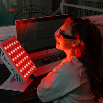
NASA research in the 1990s–2000s established near-infrared light as an effective wound healing adjunct in aerospace medicine applications, catalyzing modern LED array development. Contemporary research confirms that LED arrays at equivalent doses produce outcomes comparable to laser probes for wound healing applications, making panel-based devices clinically relevant. PBM is currently used in wound care centers as a recognized adjunct, and several systematic reviews support its incorporation into standard wound management protocols for chronic wounds.

Mechanism of Action: How PBM Affects Wound Healing

PBM accelerates wound healing through simultaneous stimulation of multiple cell types in the wound environment. In fibroblasts, photon absorption increases proliferation rate and collagen synthesis (type I and III). In keratinocytes, PBM enhances migration, differentiation, and re-epithelialization. In macrophages, PBM promotes M1→M2 polarization, shifting the wound from chronic inflammation to the proliferative repair phase. VEGF upregulation drives angiogenesis, improving blood supply and oxygen delivery to healing tissue.

  • Stimulates fibroblast proliferation and type I/III collagen synthesis — key for dermal repair
  • Enhances keratinocyte migration and re-epithelialization — accelerates wound closure
  • Promotes macrophage M1→M2 polarization, facilitating transition from inflammation to repair phase
  • Upregulates VEGF, FGF, and PDGF — growth factors driving angiogenesis and tissue proliferation
  • Increases TGF-β1 expression — promotes granulation tissue formation and remodeling
  • Enhances leukocyte activity against bacterial biofilm in wound bed
  • Improves local microcirculation via nitric oxide-mediated vasodilation
  • Reduces scar tissue formation by modulating MMP expression and collagen cross-linking

What the Research Shows: Wound Healing

Studies in this category commonly demonstrate:

  • RCTs show 30–60% faster wound closure in chronic diabetic ulcers with PBM vs. standard care alone
  • Pressure ulcer studies: significantly improved healing rates and reduced wound area versus standard care
  • Post-surgical wounds: reduced healing time and scar quality improvement with PBM
  • Animal models consistently demonstrate accelerated wound closure and improved breaking strength at closure
  • VEGF and TGF-β upregulation documented in wound tissue biopsies following PBM
  • NASA-sponsored research confirmed near-infrared LED arrays enhance wound healing in space medicine
  • Red light (630–660 nm) most effective for superficial wounds; NIR for deep or compromised wounds
  • Combination with standard wound care (moist dressings, debridement) produces superior outcomes versus either alone
  • Diabetic wound healing: NIR at 810–830 nm penetrates compromised tissue more effectively
  • Minimal side effects — PBM is safe to apply to open wound beds at therapeutic doses

Key Clinical Studies: Wound Healing

A curated selection from 450++ indexed studies.

Randomized Controlled Trial

LLLT for diabetic foot ulcer healing: double-blind RCT

Population: Human (n=68, type 2 diabetes, chronic foot ulcers)Wavelength: 830 nmDose: 4 J/cm²Year: 2013

Aziz et al. found significantly faster ulcer closure (−45% time to healing) and reduced wound area at 12 weeks in PBM group vs. standard care placebo. Demonstrated direct benefit in chronic diabetic wounds with adequate penetration via NIR.

View on PubMed →
Systematic Review & Meta-Analysis

Photobiomodulation for pressure ulcer healing: systematic review

Population: Human (8 RCTs, n=309, pressure ulcer patients)Wavelength: 630–904 nmDose: VariousYear: 2014

Systematic review found PBM significantly reduced pressure ulcer area and improved healing rates vs. control. Benefit was consistent across study designs and wound types. Supported adjunct use of PBM in pressure ulcer management protocols.

View on PubMed →
Randomized Controlled Trial

LED phototherapy accelerates post-surgical wound healing and reduces scar formation

Population: Human (n=45, elective surgery patients)Wavelength: 633 + 830 nmDose: 3 J/cm²Year: 2015

Post-surgical LED PBM (3×/week for 4 weeks) significantly reduced wound healing time, improved scar quality scores (VSS), and reduced scar erythema compared to control. Combined 633/830 nm showed superior outcomes to single wavelength.

View on PubMed →
Preclinical Study

Near-infrared LED arrays increase VEGF and TGF-β1 in wound healing (NASA-funded)

Population: Preclinical (murine excisional wound model + cell culture)Wavelength: 670 nmDose: 4 J/cm²Year: 2001

NASA-funded study by Whelan et al. found LED irradiation at 670 nm significantly increased VEGF, TGF-β1, and basic FGF in wound tissue, with accelerated closure in murine models. Formed the basis for LED array development in wound care.

View on PubMed →
Randomized Controlled Trial

LLLT for venous stasis ulcer healing: randomized controlled trial

Population: Human (n=60, chronic venous ulcers)Wavelength: 660 nmDose: 3 J/cm²Year: 2016

PBM at 660 nm (3×/week for 8 weeks) produced significantly greater reduction in venous ulcer area versus compression therapy alone. Complete healing achieved in 47% of PBM group vs. 20% in control. Established additive benefit of PBM plus standard compression.

View on PubMed →
Meta-Analysis

Low-level laser therapy for wound healing: meta-analysis of randomized controlled trials

Population: Human (12 RCTs, various chronic wound types)Wavelength: 630–904 nmDose: 1–5 J/cm²Year: 2017

Meta-analysis across 12 RCTs found PBM significantly reduced wound area (−38% vs. control) and time to healing. Effect sizes were moderate (SMD ~0.7). Identified dose range 1–4 J/cm² as most effective for superficial wound tissue.

View on PubMed →

Typical Research Parameters: Wound Healing

Based on analysis of 450++ peer-reviewed studies:

ParameterTypical RangeNotes
Wavelength 630–660 nm (red, superficial); 810–830 nm (NIR, deep/compromised) Red most effective for superficial re-epithelialization. NIR for deep wounds, diabetic tissue, or compromised vasculature.
Dose (fluence) 1–6 J/cm² Lower doses (1–4 J/cm²) effective for active wound beds. Higher doses (4–8 J/cm²) used for perilesional tissue and scar prevention.
Application method Non-contact irradiation; direct wound bed PBM can be applied directly to open wound beds at therapeutic doses without damage. Sterile technique maintained.
Session frequency 3–5× per week Most RCTs use 3× per week. Daily application used in acute post-surgical wounds. Chronic wounds: 3–5× per week until closure.
Treatment duration 4–12 weeks or until wound closure Duration tied to wound type: acute post-surgical 2–4 weeks; chronic diabetic/venous ulcers 8–16 weeks.
Supported conditions Diabetic ulcers, pressure ulcers, venous ulcers, post-surgical wounds Strongest evidence for chronic wounds. Post-surgical and post-laser wounds also studied. Burns: limited but positive preclinical data.

Frequently Asked Questions: PBM & Wound Healing

Can red light therapy accelerate wound healing?

Yes — this is one of the most evidence-supported applications of PBM. Multiple RCTs and meta-analyses confirm faster wound closure, reduced wound area, and improved tissue quality with PBM adjunct therapy versus standard care alone. Effect sizes are moderate to large for chronic wounds (diabetic ulcers, pressure ulcers, venous ulcers), where standard care often produces inadequate healing rates. Acute wounds also respond, with faster re-epithelialization and reduced scar formation.

What wavelength is best for wound healing?

Both red (630–660 nm) and near-infrared (810–830 nm) wavelengths support wound healing via different mechanisms and tissue depths. Red light primarily stimulates keratinocytes and superficial fibroblasts for re-epithelialization. NIR penetrates deeper dermal layers and is preferred for wounds with significant depth, diabetic tissue (where microvascular compromise reduces superficial light delivery), or post-radiation skin. Combined 630/830 nm protocols may offer the broadest wound coverage.

Is red light therapy safe to apply to open wounds?

At therapeutic doses, PBM is safe to apply directly to open wound beds. Therapeutic PBM irradiances (20–100 mW/cm²) are far below tissue damage thresholds, and no wound-worsening has been reported in any published human trial. Standard sterile technique is maintained during application. Contraindications include active malignancy within the treatment area and photosensitizing medications that may sensitize wound tissue.

Does red light therapy help with scar formation?

RCTs in post-surgical patients show PBM reduces scar erythema, improves scar elasticity, and lowers objective scar quality scores (VSS, POSAS) compared to controls. Mechanisms include modulation of MMP expression, regulation of collagen cross-linking, and reduction of myofibroblast activity. Treatment should ideally begin shortly after wound closure and continue for 4–8 weeks for best scar outcomes.

What is the evidence for PBM in diabetic wound healing?

Diabetic foot ulcers are a particularly well-studied application. RCTs show significantly faster closure times (−40–50% reduction in healing duration) and improved complete healing rates with NIR PBM adjunct therapy in diabetic wounds. The mechanism is especially relevant in diabetes: PBM restores mitochondrial function in metabolically compromised diabetic cells, increases NO-mediated perfusion in poorly vascularized tissue, and reduces the chronic hyperinflammatory state that impairs diabetic wound healing.

How is PBM used in wound care centers?

PBM has been incorporated as an adjunct in wound care centers and hospital wound management programs in several countries. Typical protocols use dedicated wound care laser probes or LED arrays, applied 3–5 times per week as part of comprehensive wound management (debridement, moist dressings, offloading). Evidence-based protocols are available from WALT and the North American Association for Photobiomodulation Therapy (NAALT). Panel LED devices are increasingly used as accessible alternatives to laser probes.

Browse All Wound Healing Studies

All studies in this category from the PBM research database.

Search all 10,068+ studies across all categories: Open the Full Evidence Explorer →